Reading Time: 4 Min
Dental radiography has revolutionized the way dentists diagnose and treat oral health conditions. Among the various types of dental imaging techniques, the Orthopantomogram (OPG), also known as a panoramic dental X-ray, stands out as one of the most valuable diagnostic tools in modern dentistry. This comprehensive guide will explore everything you need to know about dental OPGs, from their technical aspects to their clinical applications.
What is a Dental OPG?
An Orthopantomogram is a panoramic radiographic image that captures a complete view of the oral cavity, including all teeth, both upper and lower jaws (maxilla and mandible), temporomandibular joints (TMJ), and surrounding structures in a single two-dimensional image. This comprehensive view makes it an invaluable tool for dental diagnosis and treatment planning.

Historical Development
The journey of panoramic dental imaging began in the early 20th century, with the first patent for a panoramic X-ray machine filed by Dr. Hisatugu Numata in 1933. However, it wasn’t until the 1950s that Dr. Yrjö Paatero developed the first commercially successful panoramic X-ray machine, which he called the Orthopantomograph. Since then, the technology has evolved significantly, incorporating digital sensors and advanced imaging software.
How Does an OPG Work?
The technology behind OPG imaging is fascinating and involves several key components working in perfect synchronization:
Technical Process
- The X-ray tube and image receptor rotate around the patient’s head in a predetermined path
- The beam is collimated into a narrow vertical slit
- The receptor moves in the opposite direction to the X-ray source
- Digital sensors capture the data and convert it into a comprehensive image
- Advanced software processes the data to create a clear, detailed panoramic view
This synchronized movement allows for the capture of structures that lie in the focal trough, while blurring out structures outside this zone. The result is a clear image of the dental and maxillofacial structures.

Clinical Applications
Diagnostic Uses
Dental OPGs serve numerous diagnostic purposes in both general dentistry and specialized practice:
General Assessment
- Evaluation of tooth development and eruption patterns
- Detection of impacted teeth, particularly wisdom teeth
- Assessment of bone levels and periodontal health
- Identification of dental caries and existing restorations
Orthodontic Planning
- Analysis of dental development stages
- Assessment of tooth positioning and spacing
- Evaluation of jaw relationships
- Planning for orthodontic treatment
Surgical Planning
- Pre-surgical assessment for wisdom tooth extraction
- Implant placement planning
- Evaluation of bone quality and quantity
- Assessment of pathological conditions

Treatment Plan: Restorative work in the maxillary left quadrant, potential crown or implant consideration for the maxillary right premolar region, mandibular left restoration needs, and indicated third molar extraction in the maxillary right quadrant.

Treatment Plan: Third molar removal in the lower jaw, restoration work required in the mandibular left posterior region, and multiple restorative procedures indicated in the maxillary left quadrant

Treatment Plan: Multiple dental implants and restorative work, with additional treatment indicated including endodontic therapy in the left mandibular region and restorative procedures in both left and right lower quadrants.

Treatment Plan: Multiple extractions in both lower quadrants, endodontic therapy and restoration in the upper right region, and proposed dental implant placement in several areas of the lower jaw. The image also shows existing dental work in the upper anterior region.

Treatment Plan: A zirconia crown restoration in the lower right quadrant and multiple dental implant placements in both the right and left posterior mandibular regions. The image demonstrates successful integration of modern dental restoration techniques.
Advantages of OPG
- Comprehensive View: Captures all teeth and surrounding structures in a single image
- Patient Comfort: Quick and non-invasive procedure
- Reduced Radiation: Lower cumulative radiation dose compared to multiple individual X-rays
- Time Efficiency: Rapid image acquisition and processing
- Cost-Effectiveness: Provides extensive diagnostic information in one exposure
Limitations and Considerations
While OPGs are incredibly useful, they do have some limitations:
Technical Limitations
- Two-dimensional representation of three-dimensional structures
- Potential for geometric distortion
- Cannot provide detailed views of individual teeth
- Image quality depends on correct patient positioning
Clinical Considerations
- May not be suitable for detecting small cavities
- Some anatomical structures may overlap
- Requires supplementary imaging for detailed examination
- Not recommended for routine screening without clinical indication
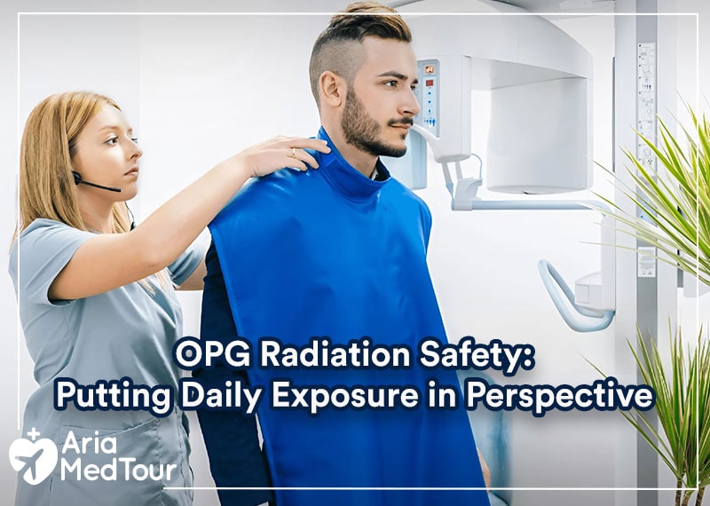
The OPG Procedure
Patient Preparation
Proper preparation and positioning are crucial for obtaining high-quality OPG images:
- Remove Metallic Objects: All removable metallic objects (jewelry, glasses, dentures) must be removed
- Position the Patient: Correct positioning in the machine using alignment lights and bite block
- Provide Instructions: Clear communication about staying still during the exposure
- Protection: Lead apron placement for radiation protection
Image Acquisition
The actual exposure process typically takes about 12-20 seconds, during which:
- The X-ray tube and detector rotate around the patient’s head
- The patient remains perfectly still
- The machine automatically adjusts exposure parameters
- Digital sensors capture the data
Dentistry Packages
Surgery + Hotel + Visa
Transfer + Interpreter
Radiation Safety and Protection
Radiation Dose
OPGs deliver a relatively low radiation dose compared to multiple individual X-rays:
- Typical effective dose: 5.5-22 μSv
- Equivalent to 1-3 days of natural background radiation
- Significantly less than a full mouth series of intraoral radiographs
Safety Measures
For Patients
- Lead apron protection
- Thyroid collar when appropriate
- Limitation of exposure frequency
- Digital imaging to minimize radiation dose
For Operators
- Distance from radiation source
- Protected positioning behind barriers
- Regular equipment maintenance and calibration
- Quality assurance programs
Digital OPG Technology
Modern Advances
Recent technological developments have significantly improved OPG imaging:
Digital Sensors
- Improved image quality
- Reduced radiation exposure
- Immediate image availability
- Digital storage and sharing capabilities
Software Enhancement
- Image manipulation tools
- Measurement capabilities
- Contrast adjustment
- Digital filters for optimal visualization
Interpretation and Analysis
Systematic Approach
Dental professionals follow a structured approach when analyzing OPG images:
- General Overview
- Image quality assessment
- Patient positioning evaluation
- Anatomical landmark identification
- Detailed Examination
- Dental structures
- Periodontal conditions
- Bone patterns
- Pathological findings
- Documentation
- Findings recording
- Treatment planning
- Patient communication
Common Pathological Findings
OPGs can reveal various pathological conditions:
- Dental caries and periapical lesions
- Periodontal bone loss
- Cysts and tumors
- Fractures and trauma
- Developmental anomalies
- TMJ disorders
You May Like to See Before & After Photos of Dentistry
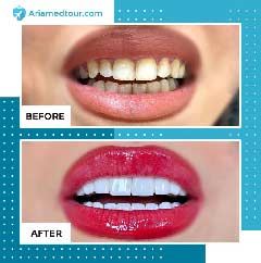
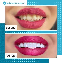
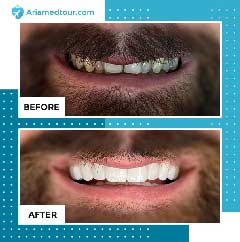
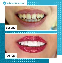
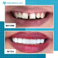
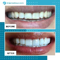
You May Like to See Before & After Photos of Dentistry
Future Developments
Emerging Technologies
The field of dental imaging continues to evolve with promising developments:
Artificial Intelligence Integration
- Automated image analysis
- Pathology detection algorithms
- Treatment planning assistance
- Quality assurance improvements

3D Capabilities
- Hybrid imaging systems
- Enhanced diagnostic accuracy
- Improved treatment planning
- Better patient communication
Conclusion
Dental OPG remains a cornerstone of modern dental diagnostics, offering an invaluable combination of comprehensive imaging, patient comfort, and diagnostic utility. As technology continues to advance, we can expect even more sophisticated applications of this essential diagnostic tool. Understanding its capabilities, limitations, and proper usage ensures optimal patient care and treatment outcomes in dental practice.
The continuous evolution of OPG technology, coupled with emerging digital innovations and artificial intelligence integration, promises to further enhance its utility in dental diagnosis and treatment planning. As we look to the future, the role of OPG in dental practice will likely become even more significant, particularly as new applications and improvements in image quality continue to develop.
Let’s Take a Look at Our Dentistry Patients’ Testimonial Videos


Related Articles
24/7 Online Support
AriaMedTour support team is available 24/7 to assist you with your inquiries about Dentistry in Iran.

Feel free to express your opinions or ask your questions regarding the article
Nice post. I learn something totally new and challenging on websites
Thank you for your comment dear… We are glad that you are reading and sharing your opinion with us
Good post! We will be linking to this particularly great post on our site. Keep up the great writing
That was good news, and thank you for sharing it with us dear.
Very well presented. Every quote was awesome and thanks for sharing the content. Keep sharing and keep motivating others.
Thank you for your comment dear… We are glad that you are reading and sharing your opinion with us
I must say this article is extremely well written, insightful, and packed with valuable knowledge that shows the author’s deep expertise on the subject, and I truly appreciate the time and effort that has gone into creating such high-quality content because it is not only helpful but also inspiring for readers like me who are always looking for trustworthy resources online. Keep up the good work and write more. i am a follower.
Thanks for your comment dear. Our effort is to clarify ambiguities and provide clear and realistic content, and we’re glad you shared your comment with us.
I just had an OPG X ray done at my dentist’s office, but I honestly didn’t understand what it shows. Can someone explain what is OPG and why it’s different from regular dental X-rays?
Hi Martha! Great question. An OPG dental scan provides a panoramic view of your entire mouth — including both jaws, all teeth, and surrounding structures. To clarify, OPG stands for Orthopantomogram, and it’s especially useful for detecting impacted teeth, jaw issues, sinus concerns, and general oral health conditions. It gives your dentist a broader picture than a standard X-ray, which focuses on just one or two teeth.